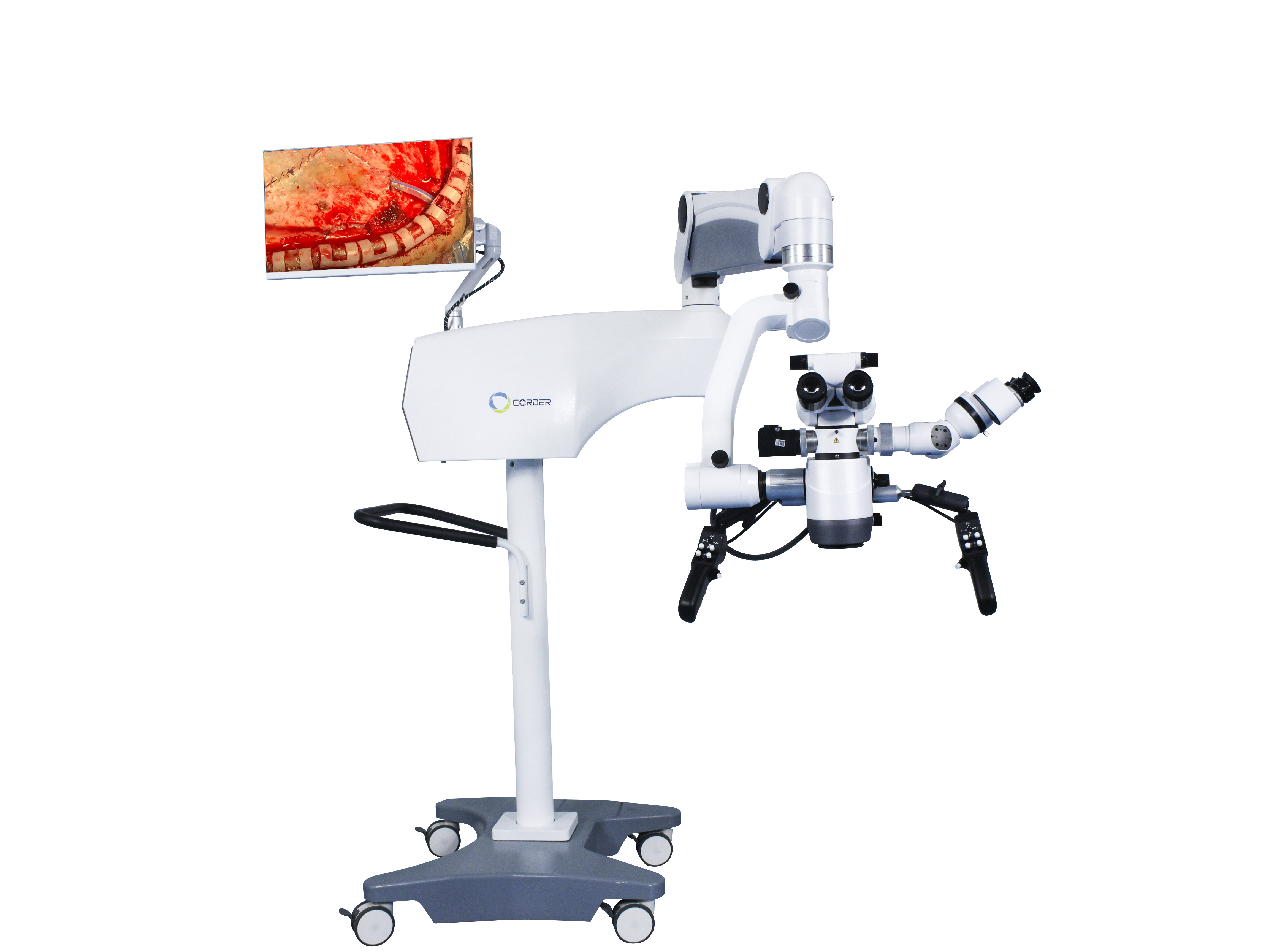
ປະຫວັດການ ນຳ ໃຊ້ແລະບົດບາດຂອງກ້ອງຈຸລະທັດຜ່າຕັດໃນການຜ່າຕັດທາງປະສາດ
ໃນປະຫວັດສາດຂອງການຜ່າຕັດ neurosurgery, ຄໍາຮ້ອງສະຫມັກຂອງກ້ອງຈຸລະທັດຜ່າຕັດເປັນສັນຍາລັກທີ່ທັນສະ ໄໝ, ກ້າວ ໜ້າ ຈາກຍຸກການຜ່າຕັດທາງປະສາດແບບດັ້ງເດີມຂອງການປະຕິບັດການຜ່າຕັດພາຍໃຕ້ຕາເປົ່າໄປສູ່ຍຸກການຜ່າຕັດປະສາດທີ່ທັນສະ ໄໝ ຂອງການປະຕິບັດການຜ່າຕັດພາຍໃຕ້ກ້ອງຈຸລະທັດ. ໃຜແລະເວລາໃດກ້ອງຈຸລະທັດປະຕິບັດການເລີ່ມໃຊ້ໃນການຜ່າຕັດທາງປະສາດບໍ? ມີບົດບາດອັນໃດກ້ອງຈຸລະທັດຜ່າຕັດມີຄົນຫຼິ້ນໃນການພັດທະນາຂອງການຜ່າຕັດ neuro? ດ້ວຍຄວາມກ້າວໜ້າຂອງວິທະຍາສາດ ແລະ ເຕັກໂນໂລຢີ, ຈະກ້ອງຈຸລະທັດປະຕິບັດການຖືກທົດແທນໂດຍອຸປະກອນທີ່ກ້າວຫນ້າທາງດ້ານຫຼາຍບໍ? ນີ້ແມ່ນຄໍາຖາມທີ່ຫມໍຜ່າຕັດ neurosurgeon ທຸກຄົນຄວນຮູ້ແລະນໍາໃຊ້ເຕັກໂນໂລຢີແລະເຄື່ອງມືຫລ້າສຸດໃນພາກສະຫນາມຂອງການຜ່າຕັດ neurosurgery, ສົ່ງເສີມການປັບປຸງທັກສະການຜ່າຕັດ neurosurgery.
1, ປະຫວັດສາດຂອງການນໍາໃຊ້ກ້ອງຈຸລະທັດໃນພາກສະຫນາມການແພດ
ໃນຟີຊິກ, ເລນແວ່ນຕາແມ່ນເລນ convex ທີ່ມີໂຄງສ້າງດຽວທີ່ມີຜົນກະທົບການຂະຫຍາຍ, ແລະການຂະຫຍາຍຂອງພວກມັນມີຈໍາກັດ, ເອີ້ນວ່າແວ່ນຂະຫຍາຍ. ໃນປີ 1590, ປະຊາຊົນຊາວໂຮນລັງສອງຄົນໄດ້ຕິດຕັ້ງແຜ່ນເລນໂຄນສອງແຜ່ນໃສ່ໃນຖັງກະບອກທີ່ຮຽວຍາວ, ດັ່ງນັ້ນຈຶ່ງໄດ້ປະດິດອຸປະກອນຂະຫຍາຍໂຄງສ້າງແບບປະສົມອັນທຳອິດຂອງໂລກ:ກ້ອງຈຸລະທັດ. ຫຼັງຈາກນັ້ນ, ໂຄງສ້າງຂອງກ້ອງຈຸລະທັດໄດ້ຖືກປັບປຸງຢ່າງຕໍ່ເນື່ອງ, ແລະການຂະຫຍາຍໄດ້ເພີ່ມຂຶ້ນຢ່າງຕໍ່ເນື່ອງ. ໃນເວລານັ້ນ, ນັກວິທະຍາສາດສ່ວນໃຫຍ່ໃຊ້ສິ່ງນີ້ກ້ອງຈຸລະທັດປະກອບເພື່ອສັງເກດເບິ່ງໂຄງສ້າງນ້ອຍໆຂອງສັດແລະພືດ, ເຊັ່ນໂຄງສ້າງຂອງຈຸລັງ. ຕັ້ງແຕ່ກາງຫາທ້າຍສະຕະວັດທີ 19, ແວ່ນຂະຫຍາຍ ແລະ ກ້ອງຈຸລະທັດ ຄ່ອຍໆຖືກນຳໃຊ້ເຂົ້າໃນຂະແໜງແພດສາດ. ໃນຕອນທໍາອິດ, ແພດຜ່າຕັດໃຊ້ແວ່ນຂະຫຍາຍແບບແວ່ນຕາທີ່ມີໂຄງສ້າງຂອງເລນດຽວທີ່ສາມາດວາງຢູ່ເທິງຂົວຂອງດັງສໍາລັບການຜ່າຕັດ. ໃນປີ 1876, ທ່ານໝໍຊາວເຢຍລະມັນ Saemisch ໄດ້ທຳການຜ່າຕັດ “ກ້ອງຈຸລະທັດ” ທຳອິດຂອງໂລກ ໂດຍໃຊ້ແວ່ນຂະຫຍາຍແວ່ນຕາປະສົມ (ບໍ່ຮູ້ຈັກປະເພດຂອງການຜ່າຕັດ). ໃນປີ 1893, ບໍລິສັດເຢຍລະມັນ Zeiss ໄດ້ປະດິດກ້ອງຈຸລະທັດ, ສ່ວນໃຫຍ່ແມ່ນໃຊ້ສໍາລັບການສັງເກດການທົດລອງຢູ່ໃນຫ້ອງທົດລອງທາງການແພດ, ເຊັ່ນດຽວກັນກັບການສັງເກດການຂອງ corneal ແລະ lesions ຫ້ອງ anterior ໃນພາກສະຫນາມຂອງ ophthalmology ໄດ້. ໃນປີ 1921, ໂດຍອີງໃສ່ການຄົ້ນຄວ້າຫ້ອງທົດລອງກ່ຽວກັບວິພາກວິພາກຂອງຫູພາຍໃນສັດ, ຜູ້ຊ່ຽວຊານດ້ານ otolaryngologist ຊູແອັດ, Nylen ໄດ້ນໍາໃຊ້ການແກ້ໄຂ.ກ້ອງຈຸລະທັດຜ່າຕັດ monocularອອກແບບແລະຜະລິດດ້ວຍຕົນເອງເພື່ອປະຕິບັດການຜ່າຕັດ otitis media ຊໍາເຮື້ອໃນມະນຸດ, ເຊິ່ງເປັນການຜ່າຕັດຈຸນລະພາກທີ່ແທ້ຈິງ. ຫນຶ່ງປີຕໍ່ມາ, ທ່ານຫມໍຊັ້ນສູງຂອງ Nylen Hlolmgren ໄດ້ແນະນໍາ aກ້ອງສ່ອງທາງໄກຜ່າຕັດຜະລິດໂດຍ Zeiss ໃນຫ້ອງປະຕິບັດການ.
ຕົ້ນກ້ອງຈຸລະທັດປະຕິບັດການມີຂໍ້ບົກຜ່ອງຫຼາຍຢ່າງ, ເຊັ່ນ: ຄວາມຫມັ້ນຄົງຂອງກົນຈັກທີ່ບໍ່ດີ, ບໍ່ສາມາດເຄື່ອນທີ່, ການສະຫວ່າງຂອງແກນທີ່ແຕກຕ່າງກັນແລະການໃຫ້ຄວາມຮ້ອນຂອງເລນຈຸດປະສົງ, ພາກສະຫນາມຂະຫຍາຍການຜ່າຕັດແຄບ, ແລະອື່ນໆ. ເຫຼົ່ານີ້ແມ່ນເຫດຜົນທັງຫມົດທີ່ຈໍາກັດການນໍາໃຊ້ທີ່ກວ້າງຂວາງ.ກ້ອງຈຸລະທັດຜ່າຕັດ. ໃນສາມສິບປີຕໍ່ໄປນີ້, ເນື່ອງຈາກການໂຕ້ຕອບໃນທາງບວກລະຫວ່າງແພດຜ່າຕັດແລະຜູ້ຜະລິດກ້ອງຈຸລະທັດ, ການປະຕິບັດຂອງກ້ອງຈຸລະທັດຜ່າຕັດໄດ້ຖືກປັບປຸງຢ່າງຕໍ່ເນື່ອງ, ແລະກ້ອງຈຸລະທັດຜ່າຕັດ, ກ້ອງຈຸລະທັດຫລັງຄາ, ເລນຊູມ, ການສ່ອງແສງແຫຼ່ງແສງສະຫວ່າງ coaxial, ເອເລັກໂຕຣນິກຫຼືນ້ໍາຄວບຄຸມແຂນ articulated, ການຄວບຄຸມ pedal ຕີນ, ແລະອື່ນໆໄດ້ຖືກພັດທະນາຢ່າງຕໍ່ເນື່ອງ. ໃນປີ 1953, ບໍລິສັດເຢຍລະມັນ Zeiss ໄດ້ຜະລິດຊຸດພິເສດກ້ອງຈຸລະທັດຜ່າຕັດສໍາລັບ otologyໂດຍສະເພາະແມ່ນເຫມາະສົມສໍາລັບການຜ່າຕັດກ່ຽວກັບບາດແຜເລິກເຊັ່ນ: ຫູກາງແລະກະດູກຂ້າງ. ໃນຂະນະທີ່ການປະຕິບັດຂອງກ້ອງຈຸລະທັດຜ່າຕັດສືບຕໍ່ປັບປຸງ, ຈິດໃຈຂອງແພດຜ່າຕັດຍັງມີການປ່ຽນແປງຢ່າງຕໍ່ເນື່ອງ. ສໍາລັບຕົວຢ່າງ, ທ່ານຫມໍເຢຍລະມັນ Zollner ແລະ Wullstein ກໍານົດວ່າກ້ອງຈຸລະທັດຜ່າຕັດຕ້ອງໄດ້ຮັບການນໍາໃຊ້ສໍາລັບການຜ່າຕັດຮູບຮ່າງເຍື່ອ tympanic. ນັບຕັ້ງແຕ່ປີ 1950, ophthalmologists ໄດ້ຄ່ອຍໆປ່ຽນການປະຕິບັດຂອງການນໍາໃຊ້ພຽງແຕ່ກ້ອງຈຸລະທັດສໍາລັບການກວດ ophthalmic ແລະແນະນໍາ.ກ້ອງຈຸລະທັດ otosurgicalເຂົ້າໄປໃນການຜ່າຕັດຕາ. ຕັ້ງແຕ່ນັ້ນມາ,ກ້ອງຈຸລະທັດປະຕິບັດການໄດ້ຖືກນໍາໃຊ້ຢ່າງກວ້າງຂວາງໃນຂົງເຂດ otology ແລະ ophthalmology.
2, ການນໍາໃຊ້ກ້ອງຈຸລະທັດໃນການຜ່າຕັດໃນ neurosurgery
ເນື່ອງຈາກພິເສດຂອງການຜ່າຕັດ neurosurgery ໄດ້, ຄໍາຮ້ອງສະຫມັກຂອງກ້ອງຈຸລະທັດໃນການຜ່າຕັດທາງປະສາດແມ່ນຊ້າກວ່າທາງດ້ານ otology ແລະ ophthalmology ເລັກນ້ອຍ, ແລະແພດຜ່າຕັດ neurosurgeons ກໍາລັງຮຽນຮູ້ເຕັກໂນໂລຢີໃຫມ່ນີ້ຢ່າງຈິງຈັງ. ໃນເວລານັ້ນ, ໄດ້ການນໍາໃຊ້ກ້ອງຈຸລະທັດຜ່າຕັດສ່ວນໃຫຍ່ແມ່ນຢູ່ໃນເອີຣົບ. ແພດຊ່ຽວຊານຕາຊາວອາເມລິກາ Perrit ໄດ້ແນະນໍາຄັ້ງທໍາອິດກ້ອງຈຸລະທັດຜ່າຕັດຈາກເອີຣົບໄປສະຫະລັດອາເມລິກາໃນປີ 1946, ວາງພື້ນຖານສໍາລັບແພດຜ່າຕັດ neurosurgeons ອາເມລິກາກ້ອງຈຸລະທັດປະຕິບັດການ.
ຈາກທັດສະນະຂອງການເຄົາລົບຄຸນຄ່າຂອງຊີວິດຂອງມະນຸດ, ເຕັກໂນໂລຢີໃຫມ່, ອຸປະກອນ, ຫຼືເຄື່ອງມືທີ່ໃຊ້ສໍາລັບຮ່າງກາຍຂອງມະນຸດຄວນໄດ້ຮັບການທົດລອງສັດເບື້ອງຕົ້ນແລະການຝຶກອົບຮົມດ້ານວິຊາການສໍາລັບຜູ້ປະກອບການ. ໃນປີ 1955, ແພດຜ່າຕັດປະສາດຂອງອາເມລິກາ Malis ໄດ້ຜ່າຕັດສະໝອງໃສ່ສັດໂດຍໃຊ້ aກ້ອງສ່ອງທາງໄກຜ່າຕັດ. Kurze, ແພດຜ່າຕັດປະສາດຂອງມະຫາວິທະຍາໄລ Southern California ໃນສະຫະລັດ, ໄດ້ໃຊ້ເວລາ 1 ປີເພື່ອຮຽນຮູ້ເຕັກນິກການຜ່າຕັດຂອງການໃຊ້ກ້ອງຈຸລະທັດຢູ່ໃນຫ້ອງທົດລອງຫຼັງຈາກສັງເກດເຫັນການຜ່າຕັດຫູພາຍໃຕ້ກ້ອງຈຸລະທັດ. ໃນເດືອນສິງຫາປີ 1957, ລາວໄດ້ປະຕິບັດການຜ່າຕັດ neuroma acoustic ສົບຜົນສໍາເລັດກັບເດັກ 5 ປີອາຍຸ 5 ປີ.ກ້ອງຈຸລະທັດຜ່າຕັດຫູເຊິ່ງເປັນການຜ່າຕັດຈຸລະພາກແຫ່ງທຳອິດຂອງໂລກ. ຫຼັງຈາກນັ້ນບໍ່ດົນ, Kurze ປະສົບຜົນ ສຳ ເລັດໃນການຜ່າຕັດເສັ້ນປະສາດຂອງເສັ້ນປະສາດຜິວ ໜັງ ພາຍໃຕ້ຜິວ ໜັງ ຂອງເດັກໂດຍໃຊ້ເຄື່ອງປັ້ນດິນເຜົາ.ກ້ອງຈຸລະທັດຜ່າຕັດ, ແລະການຟື້ນຕົວຂອງເດັກແມ່ນດີເລີດ. ນີ້ແມ່ນການຜ່າຕັດຈຸລະພາກຄັ້ງທີສອງໃນໂລກ. ຫຼັງຈາກນັ້ນ, Kurze ໄດ້ໃຊ້ລົດບັນທຸກເພື່ອຂົນສົ່ງກ້ອງຈຸລະທັດປະຕິບັດການໄປສະຖານທີ່ຕ່າງໆສໍາລັບການຜ່າຕັດ neurosurgical microsurgery, ແລະແນະນໍາໃຫ້ໃຊ້ຢ່າງແຂງແຮງກ້ອງຈຸລະທັດຜ່າຕັດກັບແພດຜ່າຕັດ neurosurgeons ອື່ນໆ. ຫຼັງຈາກນັ້ນ, Kurze ປະຕິບັດການຜ່າຕັດເສັ້ນປະສາດສະຫມອງອັກເສບໂດຍໃຊ້ aກ້ອງຈຸລະທັດຜ່າຕັດ(ຫນ້າເສຍດາຍ, ລາວບໍ່ໄດ້ເຜີຍແຜ່ບົດຄວາມໃດໆ). ດ້ວຍການຊ່ວຍເຫຼືອຂອງຄົນເຈັບທີ່ເປັນໂຣກ neuralgia trigeminal ທີ່ລາວໄດ້ຮັບການປິ່ນປົວ, ລາວໄດ້ສ້າງຕັ້ງຫ້ອງທົດລອງ neurosurgery ຖານກະໂຫຼກຫົວຫົວຈຸລະພາກຂອງໂລກຄັ້ງ ທຳ ອິດໃນປີ 1961. ພວກເຮົາຄວນຈື່ໄວ້ສະ ເໝີ ການປະກອບສ່ວນຂອງ Kurze ເຂົ້າໃນການຜ່າຕັດຈຸລະພາກແລະຮຽນຮູ້ຈາກຄວາມກ້າຫານຂອງລາວທີ່ຈະຍອມຮັບເຕັກໂນໂລຢີແລະແນວຄວາມຄິດໃຫມ່. ຢ່າງໃດກໍຕາມ, ຈົນກ່ວາຕົ້ນຊຸມປີ 1990, ບາງແພດຜ່າຕັດ neurosurgeons ໃນປະເທດຈີນບໍ່ຍອມຮັບກ້ອງຈຸລະທັດປະສາດສໍາລັບການຜ່າຕັດ. ນີ້ບໍ່ແມ່ນບັນຫາກັບກ້ອງຈຸລະທັດປະສາດຕົວຂອງມັນເອງ, ແຕ່ບັນຫາກັບຄວາມເຂົ້າໃຈຂອງ neurosurgeons' ideological.
ໃນປີ 1958, ໝໍຜ່າຕັດປະສາດຂອງອາເມລິກາ Donaghy ໄດ້ສ້າງຕັ້ງຫ້ອງວິໄຈ ແລະ ຝຶກອົບຮົມການຜ່າຕັດຈຸລະພາກແຫ່ງທຳອິດຂອງໂລກໃນເມືອງ Burlington, Vermont. ໃນໄລຍະຕົ້ນ, ລາວຍັງພົບກັບຄວາມສັບສົນແລະຄວາມຫຍຸ້ງຍາກທາງດ້ານການເງິນຈາກຜູ້ບັນຊາການຂອງລາວ. ໃນວິຊາການ, ລາວສະເຫມີຈິນຕະນາການຕັດເສັ້ນເລືອດ cortical ເປີດໂດຍກົງເພື່ອສະກັດ thrombi ຈາກຄົນເຈັບທີ່ມີ thrombosis ສະຫມອງ. ດັ່ງນັ້ນລາວໄດ້ຮ່ວມມືກັບແພດຜ່າຕັດເສັ້ນເລືອດ Jacobson ໃນການຄົ້ນຄວ້າກ່ຽວກັບສັດແລະທາງດ້ານການຊ່ວຍ. ໃນເວລານັ້ນ, ພາຍໃຕ້ເງື່ອນໄຂຂອງຕາເປົ່າ, ພຽງແຕ່ເສັ້ນເລືອດຂະຫນາດນ້ອຍທີ່ມີເສັ້ນຜ່າກາງ 7-8 ມິນລິແມັດຫຼືຫຼາຍກວ່ານັ້ນສາມາດຖືກມັດ. ເພື່ອບັນລຸ anastomosis ສຸດທ້າຍຂອງເສັ້ນເລືອດທີ່ລະອຽດກວ່າ, Jacobson ທໍາອິດໄດ້ພະຍາຍາມໃຊ້ແວ່ນຂະຫຍາຍແບບແວ່ນຕາ. ຫຼັງຈາກນັ້ນບໍ່ດົນ, ລາວຈື່ຈໍາການນໍາໃຊ້ກ້ອງຈຸລະທັດຜ່າຕັດ otolaryngologyສໍາລັບການຜ່າຕັດໃນເວລາທີ່ລາວເປັນແພດຫມໍທີ່ຢູ່ອາໄສ. ດັ່ງນັ້ນ, ດ້ວຍການຊ່ວຍເຫຼືອຂອງ Zeiss ໃນປະເທດເຢຍລະມັນ, Jacobson ໄດ້ອອກແບບກ້ອງຈຸລະທັດຜ່າຕັດຄູ່ (Diploscope) ສໍາລັບ anastomosis vascular, ເຊິ່ງອະນຸຍາດໃຫ້ຜ່າຕັດສອງຄົນເຮັດການຜ່າຕັດພ້ອມກັນ. ຫຼັງຈາກການທົດລອງສັດຢ່າງກວ້າງຂວາງ, Jacobson ຈັດພີມມາບົດຄວາມກ່ຽວກັບ microsurgical anastomosis ຂອງຫມາແລະເສັ້ນເລືອດແດງທີ່ບໍ່ແມ່ນ carotid (1960), ອັດຕາ patency 100% ຂອງ vascular anastomosis. ນີ້ແມ່ນເອກະສານທາງການແພດພື້ນຖານທີ່ກ່ຽວຂ້ອງກັບການຜ່າຕັດລະບົບປະສາດຈຸນລະພາກແລະການຜ່າຕັດເສັ້ນເລືອດ. Jacobson ຍັງໄດ້ອອກແບບເຄື່ອງມືຜ່າຕັດຈຸນລະພາກຫຼາຍຢ່າງເຊັ່ນ: ມີດຕັດຈຸນລະພາກ, ຕົວຍຶດເຂັມຈຸລະພາກ ແລະ ມືຈັບເຄື່ອງມືຈຸລະພາກ. ໃນປີ 1960, Donaghy ໄດ້ປະຕິບັດຢ່າງສໍາເລັດຜົນການຜ່າຕັດເສັ້ນເລືອດຕັນໃນສະຫມອງພາຍໃຕ້ການ.ກ້ອງຈຸລະທັດຜ່າຕັດສໍາລັບຄົນເຈັບທີ່ມີ thrombosis ສະຫມອງ. Rhoton ຈາກສະຫະລັດໄດ້ເລີ່ມການສຶກສາວິພາກວິພາກຂອງສະຫມອງພາຍໃຕ້ກ້ອງຈຸລະທັດໃນປີ 1967, ເປັນບຸກເບີກພາກສະຫນາມໃຫມ່ຂອງ microsurgical anatomy ແລະເຮັດໃຫ້ການປະກອບສ່ວນທີ່ສໍາຄັນໃນການພັດທະນາຂອງ microsurgery. ເນື່ອງຈາກຂໍ້ໄດ້ປຽບຂອງກ້ອງຈຸລະທັດຜ່າຕັດແລະການປັບປຸງເຄື່ອງມື microsurgical, surgeons ຫຼາຍແລະຫຼາຍມັກໃຊ້ກ້ອງຈຸລະທັດຜ່າຕັດສໍາລັບການຜ່າຕັດ. ແລະໄດ້ຈັດພີມມາຫຼາຍບົດຄວາມທີ່ກ່ຽວຂ້ອງກ່ຽວກັບຂັ້ນຕອນ microsurgical.
3, ການນໍາໃຊ້ກ້ອງຈຸລະທັດການຜ່າຕັດໃນການຜ່າຕັດ neurosurgery ໃນປະເທດຈີນ
ໃນຖານະເປັນຄົນຮັກຊາດຈີນຢູ່ຕ່າງປະເທດໃນຍີ່ປຸ່ນ, ອາຈານ Du Ziwei ໄດ້ບໍລິຈາກພາຍໃນປະເທດຄັ້ງທໍາອິດກ້ອງຈຸລະທັດ neurosurgicalແລະທີ່ກ່ຽວຂ້ອງເຄື່ອງມື microsurgicalໄປຫາພະແນກການຜ່າຕັດທາງປະສາດຂອງໂຮງໝໍສາຂາວິທະຍາໄລການແພດຊູໂຈວ (ປະຈຸບັນແມ່ນພະແນກການຜ່າຕັດທາງປະສາດຂອງໂຮງໝໍມະຫາວິທະຍາໄລຊູໂຈວສາຂາທຳອິດ) ໃນປີ 1972. ຫຼັງຈາກກັບຄືນໄປປະເທດຈີນ, ລາວໄດ້ທຳການຜ່າຕັດຈຸນລະພາກເປັນຄັ້ງທຳອິດ ເຊັ່ນ: ໜິ້ວທາງໃນສະໝອງ ແລະເຍື່ອຫຸ້ມສະໝອງອັກເສບ. ຫຼັງຈາກການຮຽນຮູ້ກ່ຽວກັບການມີກ້ອງຈຸລະທັດ neurosurgicalແລະເຄື່ອງມືຜ່າຕັດຈຸລະພາກ, ສາສະດາຈານ Zhao Yadu ຈາກພະແນກ Neurosurgery ຂອງໂຮງຫມໍ Yiwu ປັກກິ່ງໄດ້ໄປຢ້ຽມຢາມອາຈານ Du Ziwei ຈາກວິທະຍາໄລການແພດ Suzhou ເພື່ອສັງເກດການນໍາໃຊ້ຂອງ.ກ້ອງຈຸລະທັດຜ່າຕັດ. ສາດສະດາຈານ Shi Yuquan ຈາກໂຮງຫມໍ Shanghai Huashan ໄດ້ໄປຢ້ຽມຢາມພະແນກຂອງສາດສະດາຈານ Du Ziwei ເປັນສ່ວນຕົວເພື່ອສັງເກດຂັ້ນຕອນການຜ່າຕັດຈຸນລະພາກ. ດັ່ງນັ້ນ, ຄື້ນຂອງການນໍາ, ການຮຽນຮູ້, ແລະຄໍາຮ້ອງສະຫມັກຂອງກ້ອງຈຸລະທັດປະສາດໄດ້ຖືກກະຕຸ້ນຢູ່ໃນສູນຜ່າຕັດ neurosurgery ທີ່ສໍາຄັນໃນປະເທດຈີນ, ເຊິ່ງເປັນຈຸດເລີ່ມຕົ້ນຂອງການຜ່າຕັດລະບົບປະສາດຈຸນລະພາກຂອງຈີນ.
4, ຜົນກະທົບຂອງການຜ່າຕັດຈຸລະພາກ
ເນື່ອງຈາກການນໍາໃຊ້ຂອງກ້ອງຈຸລະທັດ neurosurgical, ການຜ່າຕັດທີ່ບໍ່ສາມາດປະຕິບັດດ້ວຍຕາເປົ່າກາຍເປັນຄວາມເປັນໄປໄດ້ພາຍໃຕ້ເງື່ອນໄຂຂອງການຂະຫຍາຍ 6-10 ເທື່ອ. ສໍາລັບຕົວຢ່າງ, ການປະຕິບັດການຜ່າຕັດເນື້ອງອກ pituitary ຜ່ານ sinus ethmoidal ໄດ້ຢ່າງປອດໄພສາມາດກໍານົດແລະເອົາເນື້ອງອກ pituitary ອອກໄດ້ຢ່າງປອດໄພໃນຂະນະທີ່ປົກປ້ອງຕ່ອມ pituitary ປົກກະຕິ; ການຜ່າຕັດທີ່ບໍ່ສາມາດປະຕິບັດດ້ວຍຕາເປົ່າສາມາດກາຍເປັນການຜ່າຕັດທີ່ດີກວ່າ, ເຊັ່ນ: tumors ສະຫມອງແລະ tumor intramedullary cord. ນັກວິຊາການ Wang Zhongcheng ມີອັດຕາການຕາຍ 10.7% ສໍາລັບການຜ່າຕັດເສັ້ນປະສາດສະຫມອງກ່ອນທີ່ຈະໃຊ້ກ້ອງຈຸລະທັດ neuroscope. ຫຼັງຈາກການນໍາໃຊ້ກ້ອງຈຸລະທັດໃນປີ 1978, ອັດຕາການຕາຍຫຼຸດລົງເຖິງ 3.2%. ອັດຕາການເສຍຊີວິດຂອງການຜ່າຕັດ arteriovenous malformation ສະຫມອງໂດຍບໍ່ມີການນໍາໃຊ້ aກ້ອງຈຸລະທັດຜ່າຕັດແມ່ນ 6.2%, ແລະຫຼັງຈາກ 1984, ດ້ວຍການນໍາໃຊ້ aກ້ອງຈຸລະທັດ neurosurgery, ອັດຕາການຕາຍຫຼຸດລົງເຖິງ 1.6%. ການນໍາໃຊ້ຂອງກ້ອງຈຸລະທັດ neuroscopeອະນຸຍາດໃຫ້ປິ່ນປົວເນື້ອງອກ pituitary ໂດຍຜ່ານວິທີການ transsphenoidal transnasal invasive ຫນ້ອຍທີ່ສຸດໂດຍບໍ່ມີການຕ້ອງການ craniotomy, ຫຼຸດຜ່ອນອັດຕາການຕາຍຂອງການຜ່າຕັດຈາກ 4.7% ເປັນ 0.9%. ຜົນສໍາເລັດຂອງຜົນໄດ້ຮັບເຫຼົ່ານີ້ແມ່ນເປັນໄປບໍ່ໄດ້ພາຍໃຕ້ການຜ່າຕັດຕາລວມຍອດແບບດັ້ງເດີມ, ດັ່ງນັ້ນກ້ອງຈຸລະທັດຜ່າຕັດເປັນສັນຍາລັກຂອງການຜ່າຕັດ neurosurgery ທີ່ທັນສະໄຫມແລະໄດ້ກາຍເປັນຫນຶ່ງໃນອຸປະກອນການຜ່າຕັດທີ່ຂາດບໍ່ໄດ້ແລະບໍ່ສາມາດທົດແທນໄດ້ໃນການຜ່າຕັດ neurosurgery ທີ່ທັນສະໄຫມ.

ເວລາປະກາດ: ວັນທີ 09-09-2024


